Macromolecular Crystallography is a technique used to study biological molecules such as proteins, viruses and nucleic acids (RNA and DNA) to a resolution higher than ~5Å. This high resolution helps elucidate the detailed mechanism by which these macromolecules carry out their functions in living cells and 
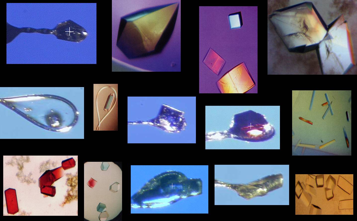
organisms. Protein molecules can crystallize under regulated conditions; the crystals are made up of multiple copies of the molecule arranged in 
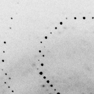
a regular 3-dimensional lattice. The x-rays deflected ("scattered") by the atoms in equivalent positions in the crystal lattice concentrate into sharp intense spots (crystal diffraction pattern). The macromolecular structure can be determined by analysis of the intensities and positions of the diffraction spots.

The Macromolecular Crystallography Group at the Stanford Synchrotron Radiation Lightsource operates and develops beamlines providing state of the art macromolecular crystallography facilities and support for visiting researchers. Of the beamlines currently operational BL12-1 and BL12-2, with an undulator source, are optimized for microfocus applications, but can also be used for conventional experiments (MAD, screening, etc.), while BL9-2 and BL14-1 are designed for MAD experiments. Researchers from universities, industry, and government laboratories around the world can gain access to the beamline facilities by submitting a research proposal.

Save the dates: The course RapiData 2024 on Data Collection and Structure Solving will take place from April 29 to May 4, 2024 and we are back in-person!. See the course announcement for more information. Application deadline: January 31, 2024.

For more news, updates on beamline facilities and other user information are posted here.

Beam Lines
Beam Line 12-1 12-2 9-2 14-1
Flux @500 mA (p/s)
and maximum beam size
4.0x10¹²
Wavelength 1.14Å
4.0x10¹²
Wavelength 0.98Å
6.4x10¹¹
Wavelength 0.98Å
1.7x10¹¹
Wavelength 1.20Å
Experiment Monochromatic,
MAD/SAD, Microbeam
Monochromatic,
MAD/SAD, Microbeam
Monochromatic,
MAD/SAD
Monochromatic,
MAD/SAD
Special instrumentation RT remote access UV-Vis microspec
Wavelength (Å)
Energy Range (keV)
0.69-2.00
6.2-18.0
0.73-1.85
6.69-17.0
0.86-2.06
6.0-14.0
0.95-2.06
6.0-13.0
Beam Size, V:H (µm) 5:40-200:150 15:15, 15:50-150:150 75:100-100:200 50:50-80:200
Detector EIGER 2XE 16M PAD EIGER 2XE 16M PAD PILATUS 6M PAD PILATUS 6M PAD
Detector Size (mm2) 311x328 311x328 431x448 431x448
Xtal-Detector (mm) 160-814 150-900 169-650 160-500
BL phone (+1-650) 926-2012 926-5212 926-5292 926-5141
Support phone (+1-650) 714-9405 714-9414 714-9417 307-4151

Common Facilities
Remote Access Remote access tools Sample Database
Remote Unix Desktop
Beamline Equipment Stanford Auto-Mounter (SAM) Xe/Kr Incubation UV-Vis microspectrophotometer Other Standard Equipment
Computing Resources Control Software Blu-Ice Computing Overview Supported Software Data Backup
Participating Research Institutions
Scripps Research Institute Stanford University

Funding