Microscopes and Video
Microscopes
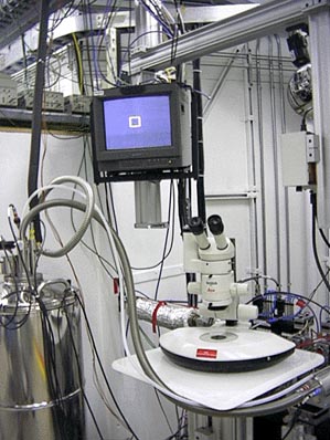
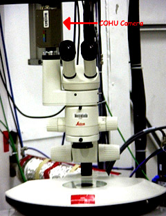
Each beamline is equipped with two Leica microscopes to aid in crystal
selection and mounting. Using a COHU camera series 2222-1340/0000 (COHU Inc. Electronics Division (858) 277-6700)
or Sony camera, one of these microscopes is setup to display images on a nearby computer monitor
and in the
beamline video system.
Directions for remote viewing:
- Focus the microscope looking through the eye pieces as usual.
- Turn on the attached video monitor or connect to the beamline video
system through a web browser.
- Flip the switch on the side of the microscope from 'vis' to 'photo'. You may lose vision through one of the eye pieces.
|
|
 |
Microscope Vendors:
The microscope is available through Lieca.
|
- Leica Inc.
111 Deer Lake Road
Deerfield, IL 60015
Phone: (800)248-0665, Fax: (847) 317-7268
|
The microscope is composed of the following parts:
|
Description (range 6.3-40X) |
Part Number |
-
M76
Optics (0.63X-4.0X Zoom)
-
Video
Phototube HV for 50%/50% View
-
0.5X
C-Mount Adapter
- 10X
C-Mount Adapter
- 45
Degree Inclined Binocular Head
-
10x Wide
Field Eyepiece
-
Reticule, 120 division w/ Crosshair
-
1.0X Achromatic Objective
-
Transmitted Light Base 20W
-
Coarse Focus Mechanism. 300 mm
-
MZ
Microscope Carrier
-
Polarizer on Stage Plate, 120 mm diam.
-
Analyzer. Rotatable. 58mm
-
Dust
Cover
|
-
10445614
-
10446194
-
D50MZC
-
D10DMC
-
10445619
-
10445111
-
10376119
-
10411589
-
10445387
-
10445615
-
10445617
-
10446228
-
10315306
-
10362677
|
| Items needed for
increased zoom (12.8-160X): |
|
-
2 ea
Eyepiece 16X/14B adjustable
-
Coarse/fine focus
drive
-
Analyzer 66mm
diameter
-
Objective Planapo
10x,M-series
-
ErgoWodge 5-25
degree
|
- 10445301
- 10447106
- 10367929
- 10446230
- 10446123
|
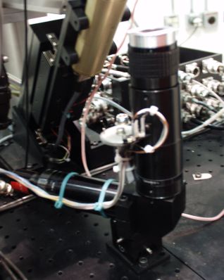
Crystal viewing in the Experimental Hutch
The sample crystal is viewed via a CCD camera and displayed in the hutch and the electronics rack with a
Samsung 170 mp / 17" flat panel monitor. The Optronics Model LE-470 Color CCD remote head camera with 6' composite and S-Video cables,
and power supply is available through Leica Inc. The sample camera uses a zoom
lens enabling the experimenter to zoom in and out while centering the crystal in
the x-ray beam. The zoom lens system is available through J.H.
Technologies in San Jose CA (408 - 436-6336). It consists of the following
components:
Navitar 12x lens with motorized zoom and manual focus, DC servo motors with encoders and optical limit switch Part# 1-50486MEL
Navitar 2X Standard NIRA Adapter Part# 1-60185
Navitar NIRA Elbow Adapter Part# 1-60165
Navitar C-Mount Coupler Part# 1-16010
Navitar 0.75X Lens Attachment, 107 mm WD Part# 1-50013
We made some modifications to this system which include a custom made limit
switch bracket holding cherry DH-series ultra-micro limit swiches (available
from DigiKey). To actuate these swiches we modified the SPUR gear that
comes with the lens and installed a 2 mm dowel pin. The system also includes a
custom mounting bracket that connects the lens to the XY-stage and a custom
mounting plate for attaching the XY-stage to the experimental table. A
lens-cover to was also added to protect the lens from LN2. The lens cover
includes a FisherBrand Preimum Cover Slip (25mm x 25mm, No. 1 12-548C from
fisher Scientific 800-766-7000). The
direct beam position is marked on the monitors using a video line generator
which was purchased from World Video, Inc. (www.microimagevideo.com).
The video signal is also entered into an axis 2400+ video server (http://www.axis.com)
|